
The electroencephalogram (EEG) is a mainstay of clinical neurology and is tightly correlated with brain function, but the specific currents generating human EEG elements remain poorly specified because of a lack of microphysiological recordings. The largest event in healthy human EEGs is the K-complex (KC), which occurs in slow-wave sleep. Here, we show that KCs are generated in widespread cortical areas by outward dendritic currents in the middle and upper cortical layers, accompanied by decreased broadband EEG power and decreased neuronal firing, which demonstrate a steep decline in network activity. Thus, KCs are isolated "down-states," a fundamental cortico-thalamic processing mode already characterized in animals. This correspondence is compatible with proposed contributions of the KC to sleep preservation(suppressing cortical arousal in response to stimuli that the sleeping brain evaluates not to signal danger)and memory consolidation.
Hmm.. alertness and knowledge preservation, huh.
Department of Neurology, Epilepsy Division, Massachusetts General Hospital, Harvard Medical School, Boston, MA 02114, USA. scash@partners.org
The human K-complex represents an isolated cortical down-state.
Cash SS, Halgren E, Dehghani N, Rossetti AO, Thesen T, Wang C, Devinsky O, Kuzniecky R, Doyle W, Madsen JR, Bromfield E, Eross L, Halász P, Karmos G, Csercsa R, Wittner L, Ulbert I.
http://www.ncbi.nlm.nih.gov/pubmed/19461004
They(K-complexes) are present in the sleep of 5 month old infants, and develop with age. Between 3 and 5 years of age a faster negative component appears and continues to increase until adolescence. Another change occurs in adults: before 30 years of age their frequency and amplitude is higher than in older people particularly those over 50 years of age.[9] This parallels the decrease in other components of sleep such as sleep spindle density and delta power.
Wauquier A. (1993). Aging and changes in phasic events during sleep. Physiol Behav 54:803–6. PMID 8248360
Individuals with restless legs syndrome have increased numbers of K-complexes and these are associated with (and often precede) leg movements. Dopamine enhancing drugs such as L-DOPA that reduce leg movements do not reduce the K-complex suggesting that they are primary and the leg movements secondary to them. Failure of such drugs to reduce K-complexes in spite of reducing the leg movements has been suggested to be why patients after such treatment still continue to complain of non-restorative sleep.[12] Clonazepam is another treatment for RLS; like other benzodiazepines, it inhibits REM sleep by enhancing levels of GABA. This inhibition of REM sleep significantly decreases K-complex count, and unlike L-DOPA treatment, clonazepam studies report improvement in sleep restoration[13]. Therefore, drugs that inhibit REM sleep also decrease K-complex count.
Saletu, M. Restless legs syndrome (RLS) and periodic limb movement disorder (PLMD) acute placebo-controlled sleep laboratory studies with clonazepam. European Neuropsychopharmacology 11:2 pgs. 153-161

Your brain is most active during REM?
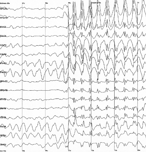
Spike-and-wave is the term that describes a particular pattern of the electroencephalogram (EEG) typically observed during epileptic seizures. Spike-and-wave patterns are seen in many types of epilepsy, and in particular during absence seizures (also called "petit mal" seizures), which are characterized by clear-cut spike-and-wave EEG oscillations. The mechanisms underlying spike-and-wave patterns are complex and involve cerebral cortex and thalamus, the intrinsic properties of the neurons, and the various types of synaptic receptors present in the circuits. In particular, it is thought that a diffuse increase in cortical excitability (either through enhanced excitation or diminished inhibition) can lead to spike-and-wave patterns through thalamocortical loops. In this case, the spike-and-wave patterns can be synchronous over large portions of the entire cerebral cortex (generalized spike-and-wave seizures). A focal increase of excitability can also entrain the thalamocortical system (or part of it) to generate spike-and-wave patterns, and various types of focal seizures.
Spike-and-wave Oscillations article in Scholarpedia


No hay comentarios:
Publicar un comentario